קובץ:GFP Superresolution Christoph Cremer.JPG
GFP_Superresolution_Christoph_Cremer.JPG (538 × 389 פיקסלים, גודל הקובץ: 156 ק"ב, סוג MIME: image/jpeg)
| זהו קובץ שמקורו במיזם ויקישיתוף. תיאורו בדף תיאור הקובץ המקורי (בעברית) מוצג למטה. |
תקציר
| תיאור |
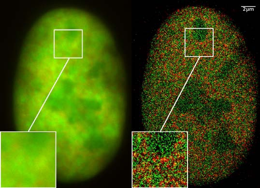
GFP superresolution, optical nanoscopy ( Christoph Cremer, emeritus at Heidelberg university [1]) View of a nucleus of a bone cancer cell: using normal high resolution fluorescence microscopy, it is not possible to distinguish details of its structure (image on the left). Using the two Color Localization Microscopy 2CLM (image on the right) it is possible to localize 70,000 histone molecules (red: RFP-H2A) and 50,000 chromatin remodeling proteins (green: GPF-Snf2H) in a field of view of 470 µm2 with an optical depth of 600 nm. Common fluorescence markers were used. 2CLM is the only optical nanoscopy method that allows position based co-localization of single molecules at high density in a wide field of view using conventional fluorescent proteins such as GFP, YFP, RFP, or other conventional fluorochromes. Due to its high optical single molecule resolution, 2CLM allows significantly more precise analyses of potential protein interactions than FRET-(Fluorescence Resonance Energy Transfer) technology, which is at present the preferred method for such investigations. This is of particular significance in studies of biomolecular machines (BMMs) within cells: Single BMMS can be analysed, including the number of molecules of a given type; distances between proteins in these BMMs often are substantially greater than those that can be analyzed by FRET (restricted to a maximum distance of only a few nm). Possible to use conventional, well established and inexpensive fluorescent dyes, from the GFP group, and its dye variants, to the well-known Alexa and fluorescein dyes. Fundamental to SPDMphymod are blinking phenomena (flashes of fluorescence), induced by reversible bleaches (metastable dark states). Individual molecules of the same spectral emission color can be detected. Publikation: Manuel Gunkel, Fabian Erdel, Karsten Rippe, Paul Lemmer, Rainer Kaufmann, Christoph Hörmann, Roman Amberger and Christoph Cremer: Dual color localization microscopy of cellular nanostructures. In: Biotechnology Journal, 2009, 4, 927-938. ISSN 1860-6768 |
| תאריך יצירה | 073009 |
| מקור | נוצר על־ידי מעלה היצירה |
| יוצר | Andy Nestl |
| אישורים והיתרים (שימוש חוזר בקובץ זה) |
|
Gallery
- Super Resolution Microscopy - Localisation Microscopy
-
 Breast Cancer Cells: 3D Dual Color Super Resolution Microscopy of Her2 and Her3 & cluster calculations
 -
 Single YFP molecule detection in a human cancer cell. Typical distance measurements 15 nm
 -
 Co- localisation microscopy with GFP and RFP fusion proteins (nucleus of a bone cancer cell) 120.000 localized molecules in a widefield area(470 µm2)
 -
 Label-free Localisation Microscopy SPDM - Super Resolution Microscopy reveals prior undetebable intracellular structures
 -
 Investigation of human eye tissue, affected by macular degeneration AMD
 -
 Virus Super Resolution Microscopy SPDM Cremer/Wege labs


רישיון
- יש לך חופש:
- לשתף – להעתיק, להפיץ ולהעביר את העבודה
- לערבב בין עבודות – להתאים את העבודה
- תחת התנאים הבאים:
- ייחוס – יש לתת ייחוס הולם, לתת קישור לרישיון, ולציין אם נעשו שינויים. אפשר לעשות את זה בכל צורה סבירה, אבל לא בשום צורה שמשתמע ממנה שמעניק הרישיון תומך בך או בשימוש שלך.
- שיתוף זהה – יצירת רמיקס, שינוי או בנייה על סמך החומר הזה, תטיל עליך חובה להפיץ את התרומות שלך לפי תנאי רישיון זהה או תואם למקור.

|
מוענקת בכך הרשות להעתיק, להפיץ או לשנות את המסמך הזה, לפי תנאי הרישיון לשימוש חופשי במסמכים של גנו, גרסה 1.2 או כל גרסה מאוחרת יותר שתפורסם על־ידי המוסד לתוכנה חופשית; ללא פרקים קבועים, ללא טקסט עטיפה קדמית וללא טקסט עטיפה אחורית. עותק של הרישיון כלול בפרק שכותרתו הרישיון לשימוש חופשי במסמכים של גנו. |
תקציר
- ↑ https://www.physik.uni-heidelberg.de/personen/lsf.php?details=1537 |titel=Fakultät für Physik und Astronomie |abruf=2020-10-01
היסטוריית הקובץ
ניתן ללחוץ על תאריך/שעה כדי לראות את הקובץ כפי שנראה באותו זמן.
| תאריך/שעה | תמונה ממוזערת | ממדים | משתמש | הערה | |
|---|---|---|---|---|---|
| נוכחית | 12:14, 30 ביולי 2009 | | 389 × 538 (156 ק"ב) | wikimediacommons>Andy Nestl | {{Information |Description=GFP superresolution, optical nanoscopy (Christoph Cremer) |Source=Own work by uploader |Date=073009 |Author=Andy Nestl |Permission=given by Christoph Cremer, University of Heidelberg |other_versions= }} |
שימוש בקובץ
הדף הבא משתמש בקובץ הזה: